


Discover super-resolution imaging with RCM
Capture datasets of 170nm resolution raw (120 after deconvolution) over a very large FOV. Study fast live-cell dynamics and perform 4D imaging in optimized conditions. The increased detection efficiency facilitates acquisition in low fluorescence conditions, like single-molecule detection (smFISH).
In combination with silicon objectives, RCM allows for deep 3D imaging of organoids, zebrafish embryos, or larger live samples.
Our solutions are as clear as our imaging
Sharper. Larger field of view. Easy to use.
Sharp images, a large field of view and easy to use? It’s possible with Confocal.nl. Get more from your samples, with a higher resolution and a higher sensitivity than conventional confocal microscopes and study live samples longer.
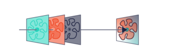
Our Re-scanning Confocal Microscope (RCM) uses multiple laser pointers and a camera as a detector. The low laser power required is live cell friendly: it prevents phototoxicity in your live samples and photobleaching of your fluorophores. Creating long-term time lapses at a super-resolution was never this accessible.
The benefits of RCM
Using RCM with a 40x 1.4 objective, you can see more cells at full resolution at once. A larger field of view increases the chances of getting the results you need.
Obtain sharp images with a high signal-to-noise ratio even in samples with a low amount of epitopes or weak stainings. Get more from your samples.
Use even lower laser power to minimise phototoxicity and photobleaching during live-cell imaging.
Getting super-resolution raw images, without averaging or integration, reduces the acquisition time and allows for a more precise analysis of the subcellular structures.
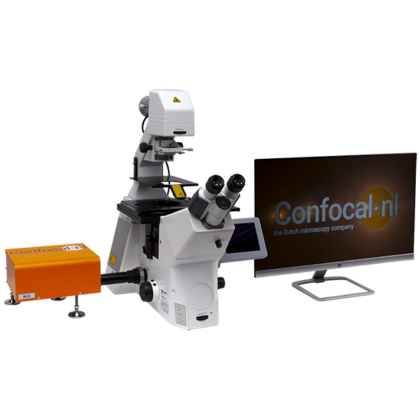
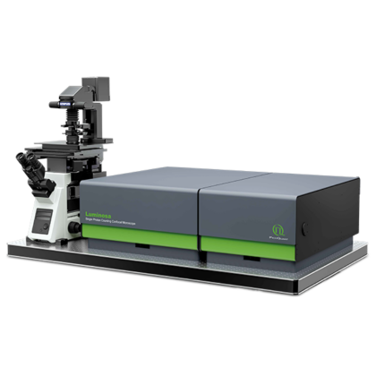
Customise your Re-Scanning confocal microscope system
- Choice of microscope bodies, e.g. Olympus IX83 – this body does an excellent job in combination with RCM. The 40x 1.4NA X-line objective gives superior image quality and FOV. If more resolution is required the 100x 1.45NA X-line is recommended.
- Hamamatsu Orca Flash 4 V3 – the high QE and low noise level of the Flash 4 V3 provide high resolution and exceptionally high SNR images in combination with RCM.
- 4 channel laser system – very stable and quiet laser engine with 405, 488, 561 and 640 laser lines.
- Tokai Hit stage top incubator – the compact Tokai Hit stage top incubator is easy to mount & remove. It is suitable for chamber slides, dishes and well plates. Happy cells guaranteed!
- LED illuminator – we like the LED light engines for their quietness and brightness. No more noisy machines and exchanging bulbs!
- Compatible with micromanager – free of charge Micromanager driver available for control of the microscope, camera & RCM. On-the-fly deconvolution by Microvolution.