


The pE-340fura provides LED illumination for Fura-2 ratiometric calcium imaging.
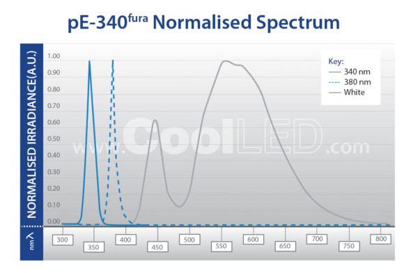
The 340nm and 380nm LED illumination system provides the optimum excitation wavelengths for Fura-2-based calcium imaging allowing high-precision, stable, high-throughput imaging with video-rate time resolution.
|
|
|
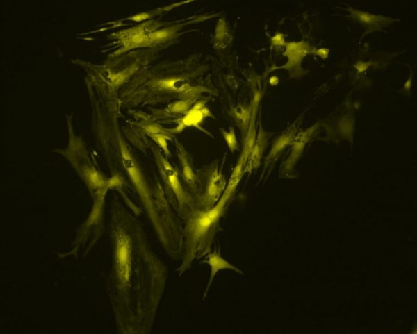
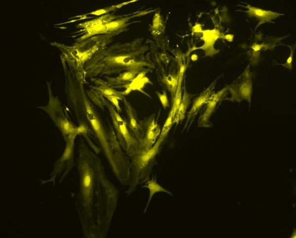
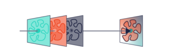
The images above show a field of cardiac myocytes (heart cells). The cells were loaded with Fura-2 using standard conditions (i.e. incubation with 2 micromolar Fura-2 acetoxymethyl ester for 30 minutes, followed by an additional 30 minutes for de-esterification.
Images were obtained by Martin Bootman and Katja Rietdorf, School of Life, Health and Chemical Sciences, The Open University, UK.
Until recently, the response time of illumination systems for Fura-2 imaging has been limited to milliseconds due to mechanical switching of the wavelengths in arc lamp and monochromator systems. However, the pE-340fura can be digitally controlled using a TTL trigger for precise illumination control in as little as 20 microseconds.
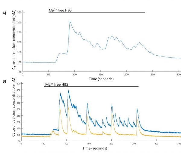
Spontaneous Ca2+ events are induced in Mg2+-free HBS. (A) Representative trace from a single hippocampal neuron of Mg2+-free induced Ca2+ events imaged at 0.5 Hz and (B) representative trace from two hippocampal neurons of Mg2+-free induced Ca2+ events imaged at 24.39 Hz.
TINNING, P. W., FRANSSEN, A. J. P.M., HRIDI, S. U., BUSHELL, T. J. and MCCONNELL, G. (2017), A 340/380 nm light-emitting diode illuminator for Fura-2 AM ratiometric Ca2+ imaging of live cells with better than 5 nM precision. Journal of Microscopy. doi:10.1111/jmi.12616
Using the pE-340fura LED illumination system for Fura-2, less dye can be loaded into the cells whilst still maintaining the same measured calcium concentration and good signal-to-noise ratio. The reduction in required dye not only improves cell-viability due to reduced dye toxicity, but also results in a cost reduction per experiment.
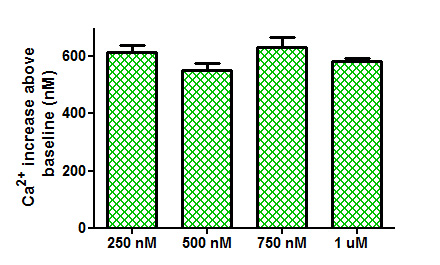
Comparison of Ca2+ increases obtained from the application of trypsin (100 nM) to tsA-201 cells loaded with different concentrations of Fura-2 AM.
TINNING, P. W., FRANSSEN, A. J. P.M., HRIDI, S. U., BUSHELL, T. J. and MCCONNELL, G. (2017), A 340/380 nm light-emitting diode illuminator for Fura-2 AM ratiometric Ca2+ imaging of live cells with better than 5 nM precision. Journal of Microscopy. doi:10.1111/jmi.12616
There are two pE-340fura configuration options:
- Direct-fit for connecting to microscopes – by selecting from a range of microscope adaptors which cover all current and most older models. A simple once only adjustment will allow optimisation to the optical path of the microscope.
- Liquid Light Guide with a fixed 3mm diameter, liquid light guide. An optional pE-340fura Universal Collimator can be specified in conjunction with a microscope adaptor if required, containing optics optimised to transmit the 340nm. CoolLED now include a light source stand for the pE-300fura illumination systems ordered with a liquid light guide (see picture 3 on the product pictures).